Somatic cell
1. Definition
Somatic cells in raw milk are usually composed of macrophages, lymphocytes, polymorphonuclear neutrophils and a small amount of breast tissue epithelial cells. The total number of somatic cells (scc) contained in each milliliter of raw milk is a quantitative indicator of its quality. And somatic cell counter can count the somatic cells in the milk. Taking raw cow milk as an example, under normal circumstances, there are about 20,000 to 200,000 individual cells per milliliter of raw cow milk. However, when the lactation system is attacked by bacteria, infected and damaged, the number of white blood cells will increase significantly to 500,000/ml to 1 million/ml or even higher.
The concentration of somatic cells is also closely related to animal husbandry management and food safety. In animal husbandry, it directly reflects the udder health and potential milk production capacity of dairy cows; in food safety, it is closely related to the quality, flavor, and elements of milk. Accurate counting of somatic cells in raw milk is very important for livestock health management and milk quality monitoring.
2. The average number of somatic cells in each country
| Country | Average Cells/ml | Year | Max. Limit Cells |
|---|
| America | 330,000 | 2008 | 750,000 |
| Canada | 260,000 | 2008 | 500,000 |
| England | 200,000 | 2004 | 400,000 |
| France | 100.000-200.000 | 2004 | 400,000 |
| The Netherlands | 210,000 | 2004 | 400,000 |
| Belgium | 203,500 | 2004 | 400,000 |
| Germany | 177,000-184,000 | 2004 | 400,000 |
| Switzerland | 139,000 | 2004 | 400,000 |
| Hungary | 250,000-260,000 | 2004 | 400,000 |
| Finland | 130,000 | 2003 | 400,000 |
| Czech | 259,000 | 2002 | 400,000 |
| Latvia | 230,000-260,000 | 2001 | 400,000 |
| China | 300,000 | 2010 | 600,000 |
What does somatic cell counter mean?
1. Definition
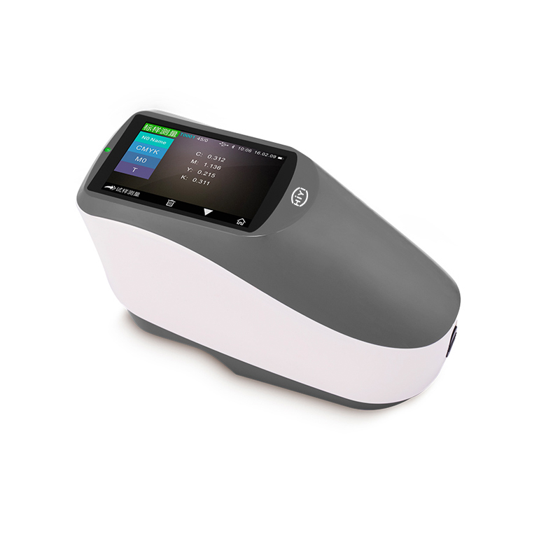
The somatic cell counter is a high-end instrument, especially for the dairy industry. It is mainly used in the quality control and product development of various dairy products, as well as the accurate counting of somatic cells in milk to improve the evaluation of the quality of liquid milk and provide quantifiable scientific data to the prevention and treatment of diseases in dairy cows in the pasture.
2. Testing indicators
The somatic cell counter for milk can count the content of somatic cells in raw milk to guide pasture management and dairy product development.
3. Scope of application
The primary tool for ranch management
In order to meet the requirements of livestock farms to improve the quality of raw milk, it is necessary to know the right information about the physiological condition of each cow. The milk yield component content and somatic cell content data obtained by this method can be used for: feeding optimization; breeding selection; early detection of subclinical mastitis.
Early detection of subclinical mastitis in dairy cows can reduce losses
Dairy cow mastitis is the most expensive dairy cow disease in the pasture. Veterinary expenses, antibiotics, retained milk, reduced production, lower milk purchase prices, and removal of sick cows, etc. The average loss per case is about $250-300.
Clinical mastitis is the abnormal milk secretion of dairy cows with the breast enlargement. Subclinical mastitis does not cause visible changes, so it is more difficult to detect. But in the subclinical mastitis stage, the milk production of dairy cows has been greatly reduced. When one cow in the herd has symptoms of clinical mastitis, 15-40 cows suffering from subclinical mastitis, which means that the potential loss caused by undetected infection is enormous.
The best way to find subclinical mastitis is to follow up the somatic cell record of each cow's milk sample month by month, just like the DHI dairy cattle breeding improvement organization.
Ensure better product quality, shelf life and profit
From the perspective of the dairy industry, the benefits of grading the purchase of raw milk based on somatic cell content are obvious, because raw milk with high somatic cell content can lead to:
A. The content of protein, fat and lactose is reduced, resulting in a decrease in cheese production, etc.
B. Increased lipase activity and free fatty acid content lead to rancidity of finished products and shortened shelf life
C. Increased levels of immunoglobulin have side effects on milk fermentation
D. The increase of sodium and chloride will produce unpleasant salt taste
4. Configuration
Automatic cleaning unit
Single sample dyeing mixer
Microprocessor control unit
Microprocessor control unit
Communication software of automatic cleaning system
Flow cytometer
Serial communication interface
With hard disk and hard drive
Parallel printer interface
Double photomultiplier
Electronic display
Flow cell measurement unit
Control software
Spare parts for 1 year
Sample detector
Sample stirrer
Printer
How do you calculate somatic cells in milk?
1. Existing detection technology
The existing somatic cell count for milk detection technology mainly targets bovine somatic cells, including indirect and direct methods.
The indirect method is mainly to detect the changes in the physical and chemical properties of milk caused by the increase of somatic cells, thereby indirectly reflecting the total number of bovine somatic cells; the direct method directly detects and counts the cells.
Common indirect methods include California Cell Count (CMT), Wisconsin Mastitis Test (WMT), viscosity method, etc. They all use surfactants to release DNA in cells and target DNA aggregates and micelles. The main drawback of these methods is that the above-mentioned physical and chemical characteristics are not typical enough to measure colloids and viscosity. Large errors will occur when the milk is not fresh, the fat content is too high, and the temperature difference is too large.
The most classic of the direct methods is the manual microscopy method, but because its operation relies too much on manual labor, it is gradually replaced by flow cytometry. The flow cytometry technology is to dilute and drive the cells that have been pre-fluorescently stained in the sheath fluid, so that they can be accurately counted one by one through the optical detection system. As the most advanced cell detection technology at present, it has fast detection speed and high accuracy, but it also has many defects, such as complicated operation process, high equipment unit price and high use cost, and it is impossible to observe cell morphology.
2. Six types of somatic cell counting methods (mainly for raw milk)
Microscopy
Add 54 ml of ethanol and 40 ml of tetrachloroethane into a 250 ml Erlenmeyer flask and shake well. Heat it in 65 °C water for 3 minutes, take it out and add 0.6 g of methylene blue, mix well, and place it in the refrigerator after cooling. After taking it out, add 6 ml of glacial acetic acid, mix well, filter with a sand core funnel, put it in a reagent bottle, and store at room temperature for later use.
The collected raw milk should be stored at 2-6° C. If it is not determined within 6 hours, add boric acid for antisepsis. The concentration of boric acid in the sample is not more than 0.6g/100mL. Store under refrigerated conditions (2-6℃) for no more than 24 hours.
Heat the raw milk in a 35 °C water bath for 5 minutes, mix it and cool it to room temperature to prevent the unevenness of the sample from interfering with the measurement results such as fat floating.
After cleaning the glass slide with ethanol, wipe it dry with lens paper, flame it to dry, and cool to room temperature.
Use microinjection to extract 0.01 ml sample, inject the sample flatly on the outer glass slide, immediately place it in the incubator, and place it horizontally for 5 minutes to form a uniform thickness sample film. Bake to dry on an electric stove, immerse the dried sample film on the slide in the staining solution for 10 minutes, take it out and air. Immerse the dyed sample membrane in water to wash away the remaining dyeing solution, and keep it dustproof after drying.
Fix the glass slide on the microscope stage, using natural light or use electric light source, condenser lens, oil immersion high power lens in order to increase the intensity of transmitted light.
The unidirectional moving mechanical stage counts the chromosomal cells on the slides in each field of view. Somatic cells that are clearly in the field of view or more than half of the shape in the field of view are used for counting, and the number of somatic cells should not be less than 400.
Microscope: magnification X500 or X1000, with graduated eyepiece, micrometer and mechanical stage;
micro syringe;
glass slide;
water bath;
electric furnace;
sand core funnel;
hair dryer;
incubator.
Reagents: 95% ethanol, tetrachloroethane or trichloroethane, methylene blue, glacial acetic acid, boric acid.
Principle
The fresh milk to be tested is smeared on a glass slide to form a film, dried, stained, and the cells whose nuclei can be clearly stained with methylene blue are counted under a microscope.
Instruments and reagents
Instruments:
Method steps
CMT
Sampling is collected from the milking area to be inspected, discard the first two milks, and squeeze the milk samples into the inspection cups of the inspection tray.
Tilt the test tray (about 60 °C), discard the excess milk, and leave about 2 ml of milk sample in the test cup.
Add 2 ml of diagnostic reagent (diluent) for dairy cows into each test cup.
Shake in horizontal concentric circles for 10-30 seconds to fully mix the milk sample with the diagnostic reagent for dairy cows.
Application
CMT is mainly used for the rapid diagnosis of latent mastitis in lactating dairy cows, the routine monitoring of breast health status of lactating dairy cows, the general survey of the incidence of latent mastitis in lactating herds, and the assessment of the hygienic quality of fresh milk.
Principle
The principle is that under the action of surface-active substances and alkaline drugs, somatic cells in the milk are destroyed and DNA is released, which further acts to cause the milk to precipitate or form a gel. The more somatic cells, the more precipitation or gel produced, which indirectly diagnoses the degree of mastitis and inflammation. This method is an internationally accepted method for the diagnosis of recessive mastitis, which is characterized by simplicity and speed.
Method
Judgment criteria
After CMT and milk are fully mixed, it is judged based on the presence or absence of agglutinates and the amount and properties of the agglutinates.
Negative (-): The number of cells is 0-200,000/ml: The mixed solution is liquid. When the test plate is tilted, the liquid moves smoothly and there is no sediment on the bottom of the cup.
Suspicious (±): The number of cells is 150,000-500,000/ml: The mixture is liquid. When the test plate is tilted, a small amount of sediment appears at the bottom of the cup, and the sediment disappears immediately after a little shake.
Weak positive (+): The number of cells is 400,000-1.5 million/ml: A small amount of thin viscous sediment appears on the bottom of the cup, but it is not gelatinous. When the test disk is tilted, the sediment is scattered on the bottom of the dish and has certain adhesion.
Positive (++): The number of cells is 800,000 to 5 million/ml: The bottom of the cup has a large amount of sediment, which is thicker and has a small amount of jelly. When the test plate is tilted, the sediment obviously adheres to the bottom of the cup. It is difficult to flow. When rotated and swayed, the sediment will be concentrated.
Strong positive (+++): The number of cells is more than 5 million/ml: Most or all of the mixture in the dish forms obvious gelatinous aggregates, which almost completely adhere to the bottom of the dish. When the test disc is rotated, the aggregates appear. The lumps are difficult to disperse.
Acidic milk (y): The milk is yellow, rancid. (mastitis)
Alkaline milk (P): The milk is dark purple, accompanied by a decrease in milk production (mastitis); when the milk is mixed with alkali, the milk tends to be dark purple.
Conductivity method
application
Mainly used to judge cattle udder disease
Principle
The conductivity of cow's milk is related to the composition, especially the chloride ion and lactose content. When the chloride ion content in milk increases or the lactose content decreases, the conductivity of the milk increases.
When the conductivity of normal milk is generally 0.004-0.005S at 20 °C. If it exceeds 0.006S, it can be considered as sick milk. Since milk contains salts, it is conductive. The number of ions in the milk will affect the conductivity, of which sodium, potassium, and chloride ions are the most influential.
When the cow's lactic acid failure or mastitis milk produces lactic acid or salt content increases, the conductivity is higher than that of normal milk. Therefore, electrical conductivity can be used to indirectly test mastitis milk.
Viscosity method
Principle
The viscosity of milk is 0.0015-0.0020 Pa.s at 20 °C. The internal factors affecting milk viscosity are the content of fat and protein in milk. If the content of fat and protein is high, the viscosity of milk is also high. The higher the dry matter content, the greater the viscosity of the milk. It is also affected by degreasing, sterilization, homogenization, etc.
At the same time, the viscosity of cow's milk is also affected by temperature. As the temperature increases, the viscosity decreases. When the temperature is lower than 60 °C, the higher the temperature, the lower the viscosity; when the temperature is higher than 60 °C, the protein denatures, and the higher the temperature, the greater the viscosity.
In addition, the viscosity of colostrum, and milk, and diseased milk are all higher than normal milk. Therefore, the viscosity can be measured to indirectly reflect the number of somatic cells.
Fluorescence photoelectric counting method
Strong operability: simple operation, convenient use, accurate results, and operators can start operation after simple training.
Strong operability: simple operation, convenient use, accurate results, and operators can start operation after simple training.
Strong operability: simple operation, convenient use, accurate results, and operators can start operation after simple training.
Supporting software: the instrument has built-in statistical software to automatically store and analyze the testing data.
Principle
The somatic cells in raw milk are fluorescently stained. After a series of lenses and filters are processed under special fluorescence, they are projected into the lens, and then the sample is photographed, read, analyzed and displayed.
Features
FCM
application
Flow cytometry (FCM) is a fast, accurate and objective technology developed in the 1970s that can simultaneously detect and quantify multiple characteristics of a single particle (usually a cell). The dairy industry mainly uses flow cytometry to detect the total number of somatic cells and bacteria in milk.
Technology
The flow cytometer is a new high-tech instrument that integrates laser technology, electronic physics technology, photoelectric measurement technology, electronic computer and cell fluorescence chemistry technology.
Structure
Its structure is generally divided into 3 parts: sample chamber and liquid flow drive system; laser light source and optical system; signal detection, storage, and data processing system.
Principle
The sample solution stained by the staining solution is pressed into the center of the nozzle through the sample tube, and the cell-free sheath solution is also pressed into the nozzle to form a sheath solution stream surrounding the cells, so that the cells enter the sample chamber in a single row, intersecting vertically with the laser beams (wavelength 488 nm) in the horizontal direction.
The stable liquid flow propulsion device makes the sample arranging in a single row, and each cell passes through the laser irradiation area in an equal time. After the fluorescently stained cells are irradiated, two signals are generated. One is the scattered light signal and the other is forward. The forward scatter (FSC) is generally proportional to the size of the cell; the side scatter (SSC) is related to the granularity and complexity of the cell.
The other is a fluorescent signal, the emitted fluorescence is directly proportional to the binding site. Different types of optical signals are received by corresponding optical detectors. The optical signal is collected by the electrical system, converted into electrical signal, and processed by software, so the detection result is obtained.
Top 10 somatic cell counter
1. LACTOS (Peru)
High-end direct fluorescent, low magnification microscope with fast autofocus and cell counting software.
Fast, accurate, reliable somatic cells analysis of cow, sheep, goat, buffalo and human milk.
Possibility of reference analysis
Detects subclinical and clinical mastitis
Lowest cost per test of Somatic Cell Counts
Easily readable results within 20 to 60 seconds
Takes a maximum of 60 images by computer-controlled X:Y movements and then processes these by the image analysis software
Stores an unlimited number of record in the database
2. CellDrop by Denovix
CellDrop Automated Cell Counters feature patented DirectPipette™ Technology to eliminate plastic slides and cumbersome hemocytometers from routine cell counting. CellDrop Cell Counters feature dual fluorescence and brightfield optics, variable height sample chamber, and powerful, easy-to-use analysis software.
3. NucleoCounter by Chemometec (Denmark)
The NucleoCounter® SCC-100™ Somatic Cell Counter is a high-quality image cytometer capable of measuring the number of somatic cells in a milk sample with high precision and accuracy.
4. LACTOSCAN SCC by LACTOSCAN (Bulgaria)
LACTOSCAN SCC Somatic cell counter is based on fluorescent image cytometry for counting cells in milk. It fully complys with the ISO 13366-1 IDF 148-1 Enumeration of somatic cells - Microscopic method (Reference method) with regard to the staining of cells, in the definition of cells to be counted depending on the size and the methodology of the counting. The somatic cell counter offers visual control for excluding accidental errors.
5. Ekomilk Horizon by EKOMILK (Bulgaria)
The automated Ekomilk Horizon ESSENTIAL is a Somatic Cell Counter using the worldwide accepted viscosity method to determine the Somatic Cell Count (SCC).
6. Mrclab (Israel)
High-end direct fluorescent, low magnification microscope with fast autofocus and cell counting software. Fast, accurate, reliable somatic cells analysis of cow, sheep, goat, buffalo and human milk.
7. Gem scientific (UK)
DeLaval Cell Counter DCC offers you a professional tool for mastitis diagnostics. With the DeLaval Cell Counter DCC you can test and monitor the somatic cell count directly on the farm and have the result in under a minute.
8. ADAM MC2 by NanoEntek (Korea)
ADAM-MC2, an automated fluorescence cell counter, performs viability and cell counting measurements using the PI (Propidium Iodide) staining method of combined with advanced image analysis. It only takes 25 seconds to get the result.
9. Corning (China)
The Corning Cell Counter generates images that are automatically analyzed using our state-of-the-art cell counting algorithm. Images are analyzed within seconds and can be used to count both mammalian cells and organoids.
10. AR BROWN (Japan)
SCC DUNK® is detectable white blood cells mainly and get the result in 3 minutes. It is applicable to an individual, or commingled cow milk samples to monitor cows with subclinical and clinical mastitis and milk quality.
Conclusion
The somatic cell counter plays an important role in controlling the quality of milk and detecting diseases in dairy cows. It is related to the actual interests of dairy farmers and milk sales and processing manufacturers. In addition, milk somatic cell counter are often used in conjunction with milk analyzers. All in all, buying the most advanced and sophisticated somatic cell counter may be the beginning of earning more profit share.
References